• Skip navigation
  • Skip to navigation
  • Skip to the bottom
Simulate organization breadcrumb open Simulate organization breadcrumb close
Chair of Visual Computing
  • FAUTo the central FAU website
  1. Friedrich-Alexander-Universität
  2. Technische Fakultät
  3. Department Informatik
Suche öffnen
  • Campo
  • StudOn
  • FAUdir
  • Jobs
  • Map
  • Help
  1. Friedrich-Alexander-Universität
  2. Technische Fakultät
  3. Department Informatik

Chair of Visual Computing

Navigation Navigation close
  • Research
    • Rendering and Visualization
    • Geometric Modeling and 3D Reconstruction
    • Virtual, Mixed, and Augmented Reality
    • Visual Computing for Digital Humanities and Social Sciences
    • Visual Healthcare Computing
    Research
  • Publications
  • Teaching
    • Vertiefungsrichtung Visual Computing
    • Summer Term 2025
    • Winter Term 2024/25
    • Theses
    Teaching
  • Staff
  • Arrival and Contact
  1. Home
  2. Research
  3. Visual Healthcare Computing

Visual Healthcare Computing

In page navigation: Research
  • Rendering and Visualization
  • Virtual, Mixed, and Augmented Reality
  • Geometric Modeling and 3D Reconstruction
  • Visual Computing for Digital Humanities and Social Sciences
  • Visual Healthcare Computing
  • Other Projects

Visual Healthcare Computing

Thomas Wittenberg

PD Dr.-Ing. Thomas Wittenberg

Department of Computer Science
Chair of Computer Science 9 (Computer Graphics)

Room: Room 01.112-128
Cauerstraße 11
91058 Erlangen
  • Phone number: +49 9131 85-25396
  • Email: thomas.wittenberg@fau.de

Office hours

Room 01.112-128, n.V.

The research area “Visual Healthcare Computing” describes the application of computer  vision, deep learning and visualization algorithms to the field of digital healthcare.
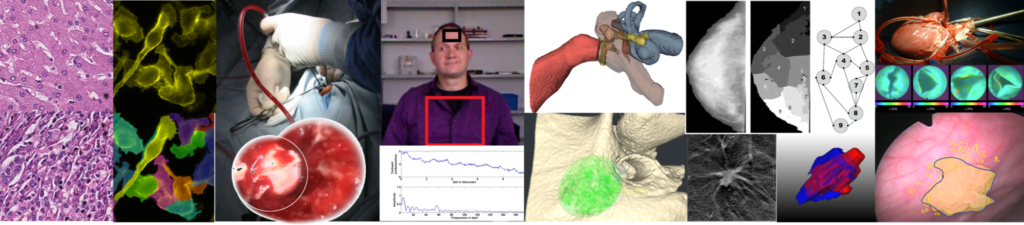
Examples are applications along a patient’s healthcare pathway, starting  from image based diagnostics (e.g. based on endoscopy or digital pathology), endoscopy-based image enhancement for orientation and navigation (using e.g. 2D/3D panoramic imaging), computer assisted radiomics (in the field of mammography), contactless monitoring of vital signs (e.g. pulse and breathing) as well as interactive immersive training and planning systems for surgical interventions.

Selected Publications:

  • Klare P., Sander C., Prinzen M., Haller M., Nowack S., Abdelhafez M., Poszler A., Brown H., Wilhelm D., Schmidt RM., von Delius S., Wittenberg T.:
    Automated polyp detection in the colorectum: a prospective study (with videos)
    In: Gastrointestinal Endoscopy 89 (2019), p. 576-582.e1
    ISSN: 0016-5107
    DOI: 10.1016/j.gie.2018.09.042
    URL: https://www.ncbi.nlm.nih.gov/pubmed/30342029
    BibTeX: Download
  • Kriegmayr M., Bergen T., Ritter M., Mandel P., Michel M., Wittenberg T., Bolenz C.:
    Digital Mapping of the Urinary Bladder: Potential for Standardized Cystoscopy Reports.
    In: Urology 104 (2017), p. 235-241
    ISSN: 0090-4295
    DOI: 10.1016/j.urology.2017.02.019
    URL: https://www.ncbi.nlm.nih.gov/pubmed/28214573
    BibTeX: Download
  • Haeberle L., Hack C., Heusinger K., Wagner F., Jud S., Uder M., Beckmann M., Schulz-Wendtland R., Wittenberg T., Fasching P.:
    Using automated texture features to determine the probability for masking of a tumor on mammography, but not ultrasound
    In: European Journal of Medical Research 22 (2017)
    ISSN: 0949-2321
    DOI: 10.1186/s40001-017-0270-0
    URL: https://www.ncbi.nlm.nih.gov/pmc/articles/PMC5577694/
    BibTeX: Download
  • Aichinger W., Krappe S., Enis AC., Cetin-Atalay R., Üner A., Benz M., Wittenberg T., Stamminger M., Münzenmayer C.:
    Automated cancer stem cell recognition in H and E stained tissue using convolutional neural networks and color deconvolution
    SPIE Medical Imaging 2017 (Orlando, Florida, USA)
    In: Proceedings Volume 10140 Medical Imaging 2017:Digital Pathology 2017
    DOI: 10.1117/12.2254036
    BibTeX: Download
  • Krappe S., Benz M., Gryanik A., Tannich E., Wegner C., Stamminger M., Wittenberg T., Münzenmayer C.:
    Automated plasmodia recognition in microscopic images for diagnosis of malaria using convolutional neural networks
    Medical Imaging: Digital Pathology 2017
    DOI: 10.1117/12.2249845
    BibTeX: Download

Chair of Visual Computing
(Lehrstuhl für Graphische Datenverarbeitung)

Cauerstraße 11
91058 Erlangen
Deutschland
  • Imprint
  • Privacy
  • Facebook
  • RSS Feed
  • Xing
Up